

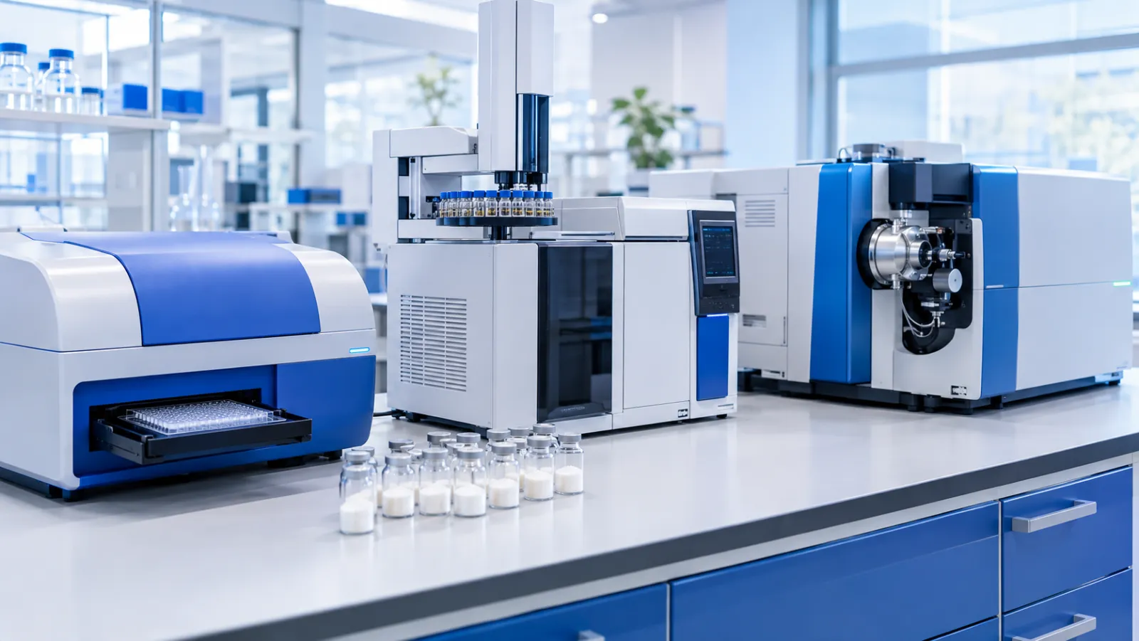
In short: Beyond identity and purity, a peptide certificate of analysis may report three contaminant tests: bacterial endotoxin (limulus or recombinant factor C assays), elemental impurities, once called heavy metals (ICP-MS), and residual solvents (headspace gas chromatography). Each follows pharmacopoeial or ICH methods, and published cell studies show how such contaminants can skew culture results.
A peptide certificate of analysis (COA) reports identity and purity first. The tests further down, for endotoxin, elemental impurities and residual solvents, look for things that are not peptide at all. They come from bacteria, from metals in reagents and equipment, and from the solvents used in solid-phase peptide synthesis. For cell-culture work, these lines deserve the same attention as the purity figure.
Why do these tests appear on a peptide COA?
They cover non-peptide impurities that synthesis and handling can leave behind. The European Medicines Agency (EMA) guideline on synthetic peptides notes that solid-phase synthesis requires extensive washing of the resin with solvents, and its example specification for medicines includes residual solvents, elemental impurities (for example where metal catalysts are used) and bacterial endotoxins 1.
The standards these tests borrow from were written for medicines. The International Council for Harmonisation (ICH) guidelines Q3C, on residual solvents, and Q3D, on elemental impurities, both state that they do not apply to products used during clinical research stages of development 2 3. When a research-grade COA quotes an ICH class or limit, it is using that scale as a reference point.
| Test | What it looks for | Common methods | Main reference texts |
|---|---|---|---|
| Bacterial endotoxins | Lipopolysaccharide from the outer membrane of Gram-negative bacteria | Limulus amebocyte lysate (gel clot, turbidimetric or chromogenic); recombinant factor C with fluorescence | USP <85> and <86>; Ph. Eur. 2.6.14 |
| Elemental impurities (“heavy metals”) | Toxic elements such as arsenic, cadmium, mercury and lead, plus catalyst metals | Inductively coupled plasma mass spectrometry (ICP-MS) | ICH Q3D; USP <232> and <233> |
| Residual solvents | Volatile organic solvents from synthesis and purification | Gas chromatography, often static headspace with flame ionisation detection | ICH Q3C; USP <467> |
What is endotoxin, and why does it matter in cell culture?
Endotoxin is lipopolysaccharide (LPS) from the outer membrane of Gram-negative bacteria 4, and some immune cells in culture respond to very small amounts of it 5. Gorbet and Sefton’s 2005 review described endotoxin as hard to avoid in the laboratory, and able to confound the effect being studied where present 6.
The studies below did not involve synthetic peptides; they show how cell readouts respond to endotoxin carried in by a reagent.
| Study | Study type | Model | What was measured | Reported finding |
|---|---|---|---|---|
| Gao, 2003 | Cell study | RAW264.7 mouse macrophage cell line | Tumour necrosis factor alpha (TNF-alpha) release with two recombinant human Hsp70 preparations of different LPS content | The less purified preparation induced TNF-alpha release; removing LPS with polymyxin B essentially eliminated it, and the authors attributed the activity entirely to contaminating LPS |
| Schwarz, 2014 | Cell study | THP-1 cells, primary human monocytes, monocyte-derived dendritic cells and primary CD1c+ dendritic cells | Response to very low LPS concentrations; endotoxin content of commercial recombinant proteins | CD1c+ dendritic cells were activated by LPS amounts equivalent to contamination found in some commercial proteins, some of which exceeded data-sheet levels |
| Uehara, 2024 | Laboratory investigation | Culture medium for oral mucosal epithelial cell sheets | Endotoxin in medium reagents, unused medium and culture supernatant | Contamination was traced to a cholera toxin supplement; the authors advised measuring reagents or confirming them with an adequate COA |
Schwarz et al. linked the dendritic cells’ sensitivity to high expression of CD14, a receptor that works with Toll-like receptor 4 (TLR4) in recognising LPS, and recommended that recombinant proteins be screened for endotoxin before work with LPS-sensitive cells 5 7.
How is endotoxin measured?
With assays built on the clotting cascade of horseshoe crab blood cells, or on a recombinant copy of its first enzyme.
Limulus amebocyte lysate (LAL). In the LAL test, endotoxin activates factor C, which activates factor B, which converts a proclotting enzyme that then clots a protein called coagulogen. The reaction can be read as a gel clot, or by turbidimetric or chromogenic methods 8.
LAL is not specific to endotoxin. Certain glucans also activate it, and commercial lysates vary in how strongly they respond to them 9. Maloney et al. traced this to a glucan-sensitive factor G in the lysate 8.
Recombinant factor C (rFC). These assays use a recombinant form of the first enzyme in the cascade and read the result by fluorescence. Because rFC lacks factor G, it is not subject to glucan false positives 8. Bolden and Smith validated an end-point fluorescence rFC method on several pharmaceutical products and reported it equivalent or superior to the compendial test 10.
Pharmacopoeial status. The European Pharmacopoeia (Ph. Eur.) has revised general chapter 2.6.14 to add the rFC fluorimetric end-point method as method G, taking over the content of chapter 2.6.32, which is to be suppressed from Issue 13.1 11. USP chapter <86> describes endotoxin tests using non-animal-derived recombinant reagents, alongside chapter <85> 12 13. The US Food and Drug Administration (FDA) accepts validated alternative methods, with the compendial gel-clot method deciding any dispute unless a monograph says otherwise 14.
Results are reported in endotoxin units (EU), read against a reference standard endotoxin such as the one USP lists for chapter <85> 12.
 Illustration: plate-based assays read endotoxin by colour, turbidity or fluorescence.
What does “heavy metals” mean on a COA?
It is an older name for what pharmacopoeias now test as elemental impurities, measured element by element.
The traditional pharmacopoeial heavy metals test was a wet-chemistry test read by visual comparison. Lewen et al. developed an ICP-MS alternative that quantified each element expected to respond in the compendial test, including arsenic, cadmium, lead, mercury, palladium and platinum, and removed the subjectiveness of the visual comparison 15.
ICH Q3D groups elements by toxicity and likelihood of occurrence, and its scope includes drug products containing synthetically produced polypeptides 3:
- Class 1: arsenic, cadmium, mercury and lead, evaluated across all sources.
- Class 2A: cobalt, nickel and vanadium, relatively likely to occur.
- Class 2B: elements including palladium, platinum, silver and gold, considered only when intentionally added, for example as catalysts.
- Class 3: barium, chromium, copper, lithium, molybdenum, antimony and tin.
Q3D names residual catalysts, manufacturing equipment and container closure systems among the sources 3. USP chapter <232> sets limits for elemental impurities in drug products, and chapter <233> describes two analytical procedures plus criteria for acceptable alternatives 16 17.
Metals matter in culture because cells respond to them. In a 2018 cell study, Keenan et al. found large differences in copper, zinc, iron, selenium and cobalt between commercial basal media, and reported that Caco-2 cell growth fell as copper rose within the range seen in those media 18.
Which residual solvents come from peptide synthesis?
Mainly the solvents used to build, wash and purify the peptide chain. Varnava and Sarojini’s 2019 review named N,N-dimethylformamide (DMF), dichloromethane and N-methyl-2-pyrrolidone (NMP) as the most common solvents in peptide synthesis 19. Acetonitrile is a usual organic component of reversed-phase HPLC mobile phases for peptides, and trifluoroacetic acid (TFA) is used for cleavage and purification 20.
ICH Q3C sorts solvents into Class 1 (to be avoided), Class 2 (to be limited) and Class 3 (low toxic potential) 2.
| Solvent | Role in peptide work | ICH Q3C listing | Q3C concentration limit |
|---|---|---|---|
| DMF | Synthesis solvent 19 | Class 2 | 880 ppm |
| Dichloromethane | Synthesis solvent 19 | Class 2 | 600 ppm |
| NMP | Synthesis solvent 19 | Class 2 | 530 ppm |
| Acetonitrile | HPLC mobile phase 20 | Class 2 | 410 ppm |
| Acetic acid | Mobile-phase additive and counter-ion 20 | Class 3 | No health-based limit; 0.5% acceptable without justification |
| Trifluoroacetic acid | Cleavage reagent and HPLC additive 20 | Solvents with no adequate toxicological data | None set |
These limits were set for medicines. ICH Q3C states that residual solvents are typically determined by gas chromatography, and that loss on drying may be used when only Class 3 solvents are present 2; USP chapter <467> covers the same ground for the United States Pharmacopeia 21. Zou et al. described a static headspace GC method with flame ionisation detection covering 27 solvents used in pharmaceutical manufacturing 22.
Solvents also register in cell assays. In a 2013 cell study, Timm et al. found that dimethyl sulfoxide (DMSO) and ethanol, both Class 3 solvents, altered interleukin-6 or reactive oxygen species readouts after LPS stimulation in some cell types, even at low percentage concentrations 23. The solvents were added deliberately as vehicles, not as residues, but culture readouts clearly responded to them.
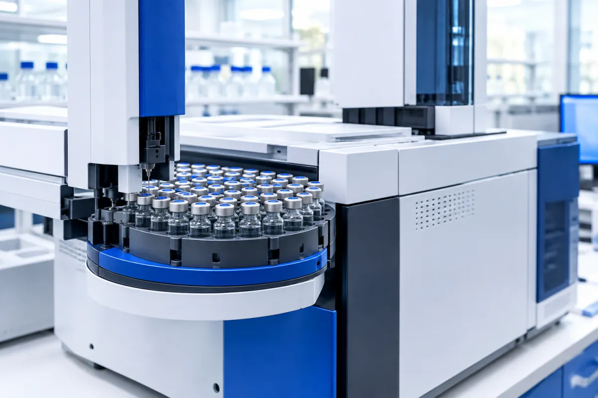 Illustration: headspace gas chromatography measures volatile solvents above a sealed sample.
How should these results be read for cell-culture work?
Check what was tested, by which method, and down to what limit.
- Endotoxin: the unit and its basis (per millilitre or per amount of peptide), the method (LAL or rFC), and whether a “less than” value is the assay’s reporting limit. The cell studies above involved macrophages, monocytes and dendritic cells.
- Elemental impurities: which elements were measured, the method, and whether numbers or only a “complies” statement are given.
- Residual solvents: which solvents were targeted. A panel that does not include DMF says nothing about DMF.
- TFA: it is not covered by ICH Q3C limits, so it appears, if at all, as counter-ion content. Our explainer on acetate vs TFA salts covers that result.
Frequently asked questions
What is an endotoxin unit (EU)?
An endotoxin unit expresses endotoxin activity measured against a reference standard endotoxin, such as the one the USP lists for its bacterial endotoxins chapter <85> 12. Results are reported per volume or per amount of sample, so a COA figure only makes sense with its basis. Maloney et al. cited one recombinant factor C assay with a range of 0.05 to 500 EU/mL 8.
What is the difference between the LAL test and recombinant factor C?
LAL is a lysate of horseshoe crab blood cells containing the whole clotting cascade, including a glucan-sensitive factor G. Recombinant factor C reproduces only the first, endotoxin-activated enzyme and reads the result by fluorescence, so glucans do not trigger it 8. The European Pharmacopoeia now includes rFC as method G in chapter 2.6.14 11, and USP chapter <86> covers recombinant reagents 13.
Why do some COAs say “elemental impurities” instead of “heavy metals”?
The older heavy metals test was a wet-chemistry test read by visual comparison. Lewen et al. described an ICP-MS alternative that quantifies each element separately and removes that subjectiveness 15. Current guidance, ICH Q3D and USP chapters <232> and <233>, sets limits and procedures for individual elements 3 16 17, so newer COAs report elements by name.
Which residual solvents might remain in a synthetic peptide?
The likeliest candidates are the solvents used in synthesis and purification. A 2019 review named DMF, dichloromethane and NMP as the most common peptide synthesis solvents 19, and acetonitrile is a usual HPLC mobile-phase component 20. ICH Q3C places all four in Class 2, the solvents to be limited 2. A useful COA lists which solvents were tested and the method, often headspace gas chromatography 22.
Is trifluoroacetic acid a residual solvent?
ICH Q3C lists trifluoroacetic acid among solvents for which no adequate toxicological data was found, so it has no Q3C limit 2. In synthetic peptides, TFA from cleavage and purification remains in cationic peptides as a counter-ion, giving trifluoroacetate salts 20, which the EMA guideline treats as a separate specification item 1. Our explainer on acetate vs TFA salts covers how it is measured.
References
- European Medicines Agency. Guideline on the Development and Manufacture of Synthetic Peptides (EMA/CHMP/CVMP/QWP/367182/2025), adopted 4 December 2025, effective 1 June 2026. https://www.ema.europa.eu/en/documents/scientific-guideline/guideline-development-manufacture-synthetic-peptides_en.pdf. Accessed 28 September 2026. [regulatory guideline]
- International Council for Harmonisation. ICH Q3C(R9) Impurities: Guideline for Residual Solvents, Step 4 version dated 24 January 2024. https://www.database.ich.org/sites/default/files/ICH_Q3C(R9)_Guideline_MinorRevision_2024_2024_Approved.pdf. Accessed 28 September 2026. [regulatory guideline]
- International Council for Harmonisation. ICH Q3D(R2) Guideline for Elemental Impurities, final version adopted 26 April 2022. https://database.ich.org/sites/default/files/Q3D-R2_Guideline_Step4_2022_0308.pdf. Accessed 28 September 2026. [regulatory guideline]
- Uehara K, Oshiro E, Ochiai A, et al. Lessons learned from contamination with endotoxin originated from the supplement in the cell culture medium. Regen Ther. 2024;27:230-233. doi:10.1016/j.reth.2024.03.022. PMID: 38596824. PMCID: PMC11002528. [laboratory investigation]
- Schwarz H, Schmittner M, Duschl A, et al. Residual endotoxin contaminations in recombinant proteins are sufficient to activate human CD1c+ dendritic cells. PLoS One. 2014;9(12):e113840. doi:10.1371/journal.pone.0113840. PMID: 25478795. PMCID: PMC4257590. [cell study]
- Gorbet MB, Sefton MV. Endotoxin: the uninvited guest. Biomaterials. 2005;26(34):6811-6817. doi:10.1016/j.biomaterials.2005.04.063. PMID: 16019062. [narrative review]
- Gao B, Tsan MF. Endotoxin contamination in recombinant human heat shock protein 70 (Hsp70) preparation is responsible for the induction of tumor necrosis factor alpha release by murine macrophages. J Biol Chem. 2003;278(1):174-179. doi:10.1074/jbc.m208742200. PMID: 12403778. [cell study]
- Maloney T, Phelan R, Simmons N. Saving the horseshoe crab: A synthetic alternative to horseshoe crab blood for endotoxin detection. PLoS Biol. 2018;16(10):e2006607. doi:10.1371/journal.pbio.2006607. PMID: 30312293. PMCID: PMC6200278. [review and validation study]
- Cooper JF, Weary ME, Jordan FT. The impact of non-endotoxin LAL-reactive materials on Limulus amebocyte lysate analyses. PDA J Pharm Sci Technol. 1997;51(1):2-6. PMID: 9099058. [narrative review]
- Bolden J, Smith K. Application of Recombinant Factor C Reagent for the Detection of Bacterial Endotoxins in Pharmaceutical Products. PDA J Pharm Sci Technol. 2017;71(5):405-412. doi:10.5731/pdajpst.2017.007849. PMID: 28733334. [method validation study]
- European Directorate for the Quality of Medicines and HealthCare (EDQM). What changes were made to general chapter 2.6.14. Bacterial endotoxins? EDQM FAQ. https://faq.edqm.eu/pages/viewpage.action?pageId=143130671. Accessed 28 September 2026. [pharmacopoeial notice]
- United States Pharmacopeia. General Chapter <85> Bacterial Endotoxins Test. USP-NF. doi:10.31003/USPNF_M98830_02_01. Accessed 28 September 2026. [pharmacopoeial chapter]
- United States Pharmacopeia. General Chapter <86> Bacterial Endotoxins Test Using Recombinant Reagents. USP-NF. doi:10.31003/USPNF_M16015_10101_01. Accessed 28 September 2026. [pharmacopoeial chapter]
- US Food and Drug Administration. Guidance for Industry: Pyrogen and Endotoxins Testing: Questions and Answers (Edition 2), March 2026. https://www.fda.gov/media/83477/download. Accessed 28 September 2026. [regulatory guidance]
- Lewen N, Mathew S, Schenkenberger M, et al. A rapid ICP-MS screen for heavy metals in pharmaceutical compounds. J Pharm Biomed Anal. 2004;35(4):739-752. doi:10.1016/j.jpba.2004.02.023. PMID: 15193718. [analytical method study]
- United States Pharmacopeia. General Chapter <232> Elemental Impurities: Limits. USP-NF. doi:10.31003/USPNF_M5192_02_01. Accessed 28 September 2026. [pharmacopoeial chapter]
- United States Pharmacopeia. General Chapter <233> Elemental Impurities: Procedures. USP-NF. doi:10.31003/USPNF_M5193_02_01. Accessed 28 September 2026. [pharmacopoeial chapter]
- Keenan J, Horgan K, Clynes M, et al. Unexpected fluctuations of trace element levels in cell culture medium in vitro: caveat emptor. In Vitro Cell Dev Biol Anim. 2018;54(8):555-558. doi:10.1007/s11626-018-0285-z. PMID: 30117037. [cell study]
- Varnava KG, Sarojini V. Making Solid-Phase Peptide Synthesis Greener: A Review of the Literature. Chem Asian J. 2019;14(8):1088-1097. doi:10.1002/asia.201801807. PMID: 30681290. [narrative review]
- Sikora K, Jaśkiewicz M, Neubauer D, et al. The Role of Counter-Ions in Peptides-An Overview. Pharmaceuticals (Basel). 2020;13(12):E442. doi:10.3390/ph13120442. PMID: 33287352. PMCID: PMC7761850. [narrative review]
- United States Pharmacopeia. General Chapter <467> Residual Solvents. USP-NF. doi:10.31003/USPNF_M99226_07_01. Accessed 28 September 2026. [pharmacopoeial chapter]
- Zou L, Guo X, McElderry JD. Platform headspace gas chromatography method for high-throughput determination of residual solvents in pharmaceutical materials. J Pharm Biomed Anal. 2023;229:115349. doi:10.1016/j.jpba.2023.115349. PMID: 36989666. [analytical method study]
- Timm M, Saaby L, Moesby L, et al. Considerations regarding use of solvents in in vitro cell based assays. Cytotechnology. 2013;65(5):887-894. doi:10.1007/s10616-012-9530-6. PMID: 23328992. PMCID: PMC3967611. [cell study]
Check the lab report
Every published certificate of analysis names its lab, lot and test date. For laboratory research use only.